Concorde Staff

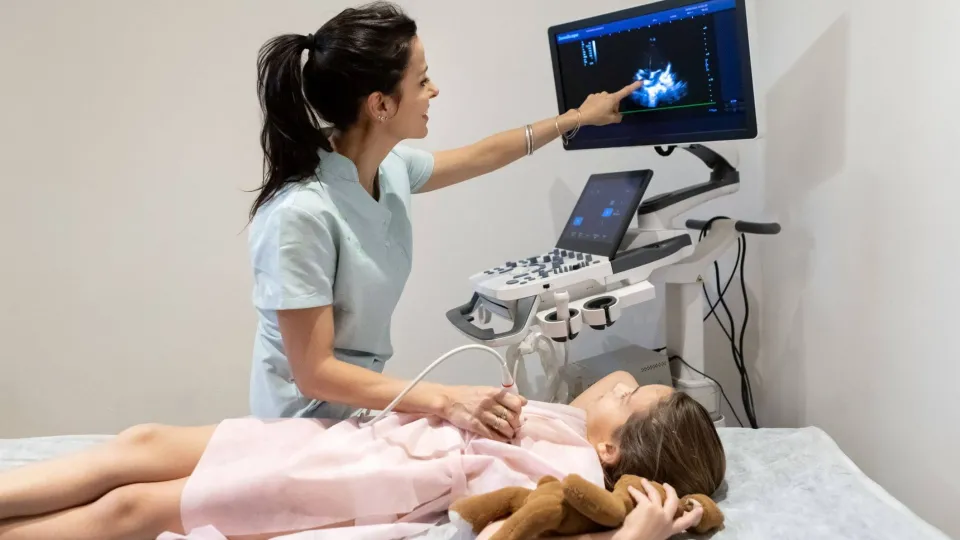
Heart disease is one of the leading causes of death in worldwide, which is why cardiac sonography is such a crucial part of the health care ecosystem. It's also an area that's especially prone to change because it centers on rapidly evolving diagnostic tools and other new technologies. Those interested in training to become a cardiac sonographer would do well to stay up to date on the current and emerging technologies in the cardiovascular ultrasound market. Here, we discuss six of the latest trends and how each one impacts the profession.
Contrast-Enhanced Ultrasound
Contrast-enhanced ultrasound, or CEUS, also known as contrast echocardiography, refers to an imaging test that incorporates an intravenous contrast agent with gas-filled microbubbles. The microbubbles flow through the blood vessels and reach the region of the heart the sonographer intends to examine. The sound waves from the cardiovascular ultrasound system cause the bubbles to vibrate and reflect the sound waves more strongly. Because blood flow is greater in areas with cancerous or diseased tissue, those areas should appear more prominently.
What CEUS means for cardiac sonography is that health care professionals may have an easier time identifying and classifying life-threatening cardiovascular diseases, such as coronary artery disease, myocardial ischemia, and hypertrophic cardiomyopathy. As this technique sees increased use in the coming years, the prognosis of cardiac events may improve thanks to early diagnosis, a key factor for desirable health outcomes.
Read more about echocardiographers: cardiac imaging experts.
3D and 4D Imaging
3D ultrasound technology involves the generation of multiple images to create a more dynamic picture, while 4D ultrasound is a live-streaming video of the 3D image. Acquiring 3D and 4D ultrasound images helps to visualize heart structures better, allows providers to study motion within the body, and may be instrumental in improving the diagnosis and management of conditions such as valvular heart disease.
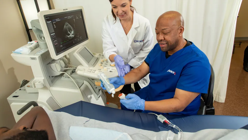
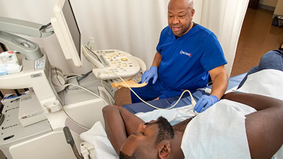
Point-of-Care Ultrasound
Point-of-care ultrasound is a technological advancement that refers to a diagnostic exam performed at the point of care — that is, at the patient's location. Point-of-care cardiovascular ultrasound systems are portable, including handheld units. Such portability allows sonographers to examine patients anywhere, the advantages of which include:
- Faster diagnosis and treatment: The sonographer can perform a diagnostic test at hospital beds or physicians' offices and get the patient to the relevant treatment as soon as possible. This practicality is crucial to emergent and near-emergent cardiac conditions.
- Broader accessibility: Point-of-care ultrasound is accessible to a broader population of patients, such as those who are too sick or injured to visit an ultrasound facility.
- Affordability: The smaller systems used in point-of-care ultrasound offer a lower cost to health care organizations and patients.
Read 10 reasons to train for a career in cardiovascular sonography.
Telecardiology
Telecardiology is an emerging trend in diagnostic imaging in the United States. It refers to the diagnosis and treatment of heart disorders using teleconferencing technology. The initial examination requires an in-person visit, but specialist interpretation, health team coordination, and subsequent patient care don't require the involved parties to be in the same location.
Telecardiology involves the use of ultrasound in a primary care setting and then transmitting the image over telecommunications lines. Specialists can then interpret the image and send back a report. The patient might also wear a watch-like electrocardiogram machine that monitors their heart condition and provides real-time data, allowing providers to respond to potentially serious events as they arise.
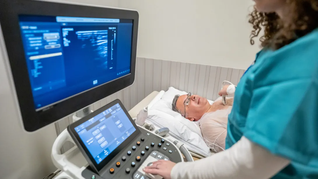
Machine Learning
Machine learning, often referred to as artificial intelligence, has seen significant growth in the cardiovascular ultrasound market and was one of the most widely discussed topics at the 2023 American Society of Echocardiography annual meeting. It has seen some use in cardiac sonography in the recent past, but research studies show that new applications continue to emerge.
For instance, the authors of a 2023 paper in the Journal of Imaging found that AI algorithms may not only help detect, classify, diagnose, and predict cardiac abnormalities but also improve the accuracy of diagnoses and speed workflow in the increasing incidence of cardiovascular diseases. That means providers may develop insights into a patient's risk factors for cardiac events and heart failure as well as the best approaches to management.
A more recent paper, published in 2024, reports on recent developments in machine learning for cardiovascular imaging, which may "add value to cardiac imaging at every step along the patient journey," including test selection, image analysis, results interpretation, diagnosis, and risk prediction for "major adverse cardiac events." These potential developments show promise for patient management and preventive medicine.
Due to the growth of the cardiovascular ultrasound system market, now is an exciting time to pursue a career in diagnostic imaging, so take the opportunity to look into the Cardiovascular Sonography Program at Concorde. You can earn your associate-level cardiovascular sonography degree in as little as 20 months and head down your career path as soon as possible. Click the "Request Info" button below or call 1-800-693-7010 to learn more about the program and our admissions process.
“The Global Burden of Cardiovascular Diseases and Risk: A Compass for Future Health,” Journal of the American College of Cardiology, https://www.jacc.org/doi/10.1016/j.jacc.2022.11.005
"3D and 4D Ultrasound: Current Progress and Future Perspectives," Current Cardiovascular Imaging Reports, https://www.ncbi.nlm.nih.gov/pmc/articles/PMC5680402
"The Role of Artificial Intelligence in Echocardiography," Journal of Imaging, https://www.mdpi.com/2313-433X/9/2/50
"Value Creation Through Artificial Intelligence and Cardiovascular Imaging: A Scientific Statement From the American Heart Association," Circulation, https://www.ahajournals.org/doi/10.1161/CIR.0000000000001202
“Cardiac Ultrasound Systems Market,” Market.Us, https://market.us/report/cardiac-ultrasound-systems-market/#:~:text=Key%20Takeaways,87%25%20of%20the%20market%20share.

Take The Next Step Towards a Brighter Future
Interested in learning more about our Cardiovascular Sonography program? We have a Concorde representative ready to talk about what matters most to you. Get answers about start dates, curriculum, financial aid, scholarships and more!